|
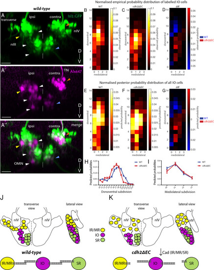
Non-<italic>cdh2ΔEC</italic> expressing zebrafish inferior oblique neurons are mispositioned following perturbation of cadherin function in neighbouring oculomotor subnuclei.(A–A’’) Transverse maximum intensity projection of a wild-type Isl1:GFP larva at 5 dpf in which nIII and nIV neurons have been labelled using retro-orbital dye fills with Alexa647 dextrans. Ipsi denotes side of dye fill. White and yellow arrows indicate ipsilateral GFP-negative IO neurons and ipsilateral GFP-positive neurons (IR/MR). Blue and orange arrows indicate contralateral GFP-positive SR neurons and SO (nIV) neurons. OMN, oculomotor nerve; TN, trochlear nerve. Scale bars = 20 μm. (B–C) Normalised empirical and posterior (E–F) probability distribution of IO neuron spatial locations in wild types (278 cells, n = 19 larvae, four clutches) and cdh2ΔEC (399 cells, n = 22 larvae, five clutches) according to dorsoventral (DV; from z0 (ventral) to z14 (dorsal)) and mediolateral (ML; from x0 (medial) to x3 (lateral)) subdivisions. (D) Normalised empirical and posterior (G) differential probability distribution of IO neuron spatial locations between genotypes. diff = cdh2ΔEC distribution – wild-type distribution. There are differences in the overall spatial distribution between genotypes (KLmean = 0.3114, 95% CI [0.3107 0.3122]). (H–I) Median posterior probabilities of IO cell location within DV (H) and ML (I) subdivisions for wild types and cdh2ΔEC larvae. There is a difference in DV (z-shift = 0.5201, 95% CI [0.5168 0.5234]) and ML distribution (x-shift = −0.1423, 95% CI [−0.1431–0.1414]) between genotypes. (J–K) Diagrammatic representation of transverse and lateral views of IO ocular motor neuron location in wild types and cdh2ΔEC. Perturbation of cadherin binding between cdh2ΔEC-expressing IR/MR and SR subnuclei and wild-type IO neurons leads to a ventral and lateral shift in IO neuron location.
|

