
Fig. 2
- ID
- ZDB-FIG-200922-2
- Publication
- Gebuijs et al., 2020 - The anti-epileptic drug valproic acid causes malformations in the developing craniofacial skeleton of zebrafish larvae
- Other Figures
- All Figure Page
- Back to All Figure Page
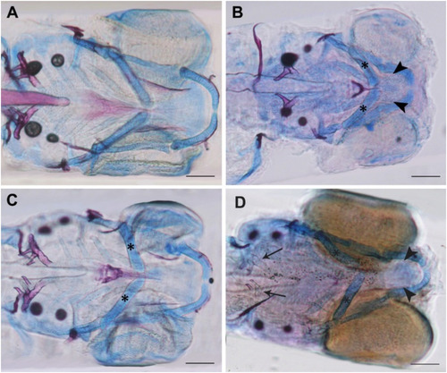
Fig. 2. A–D: Control and examples of VPA treated larvae at 5 dpf. Larvae are stained with Alcian blue (cartilage) and Alizarin red (mineralization). A: Control larvae. B: Larva treated with 100 μM VPA during 1–13 hpf, showing reduced size of cartilage elements and lacking mineralization of the notochord sheath, branchiostegal ray 1 and the entopterygoid bone (see arrowheads). C: Larva treated with 50 μM VPA during 25–37 hpf, showing small reductions in the length of the ceratohyals (asterisks). D: Larva treated with 100 μM VPA during 25–37 hpf with reduced size of cartilage elements and lacking mineralization of the notochord sheath, ceratobranchial 5 (arrows) and the entopterygoid bone (arrowheads). Scale bars are 100 μm. |
Reprinted from Mechanisms of Development, 163, Gebuijs, I.G.E., Metz, J.R., Zethof, J., Carels, C.E.L., Wagener, F.A.D.T.G., Von den Hoff, J.W., The anti-epileptic drug valproic acid causes malformations in the developing craniofacial skeleton of zebrafish larvae, 103632, Copyright (2020) with permission from Elsevier. Full text @ Mech. Dev.