
Fig. 9
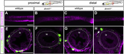
Disrupted smooth muscle development contributes to intestinal dysgenesis in dnmt1 mutants. Confocal images of 5 dpf intestines labeled with smooth muscle myosin (SMM) and desmin (magenta) antibodies. Dissected intestines (A–D) of wildtype (A,C) and dnmt1 mutants (B,D). In both proximal (B) and distal (D) intestine, dnmt1 mutants essentially lack smooth muscle cells, as revealed by whole-mount antibody staining. Transverse sections of wildtype (E,G) and dnmt1 mutants (F,H) showing loss of intestinal smooth muscle in mutants. White arrow points to phox2b:EGFP (green) positive ENS neuron that is further removed from the intestinal epithelium in mutants (H) than in wildtypes (G). ie = intestinal epithelium. Scale bar = 50 μm in A-D, and 25 μm in E-H. |
| Fish: | |
|---|---|
| Observed In: | |
| Stage: | Day 5 |
Reprinted from Developmental Biology, 455, Ganz, J., Melancon, E., Wilson, C., Amores, A., Batzel, P., Strader, M., Braasch, I., Diba, P., Kuhlman, J.A., Postlethwait, J.H., Eisen, J.S., Epigenetic factors Dnmt1 and Uhrf1 coordinate intestinal development, 473-484, Copyright (2019) with permission from Elsevier. Full text @ Dev. Biol.