
Fig. 3
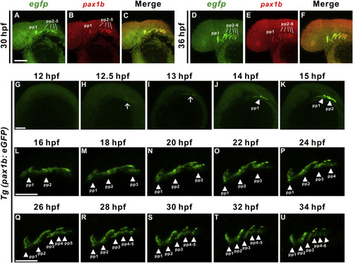
Time lapse analyses of Tg(pax1b:eGFP) enhancer trap transgenic embryos reveals sequential segmental development of pharyngeal pouches. Double fluorescence in situ hybridization indicated co-expression of egfp (A, D, green) with pax1b (B, E, red) in pharyngeal pouches (pp) 1–5 or 1–6 (C, F, merge) at 30 and 36 hpf. Time lapse analyses of Tg(pax1b:eGFP) transgenic embryos show morphogenesis of pharyngeal pouch 1–6 during 12 to 34 hpf stages. Lateral view of a Tg(pax1b:eGFP) embryo at 12 hpf (G), 12.5 hpf (H), 13 hpf (I), 14 hpf (J), 15 hpf (K), 16 hpf (L), 18 hpf (M), 20 hpf (N), 22 hpf (O), 24 hpf (P), 26 hpf (Q), 28 hpf (R), 30 hpf (S), 32 hpf (T) and 34 hpf (U) are shown. Scale bars represent 100 μm. |
| Genes: | |
|---|---|
| Fish: | |
| Anatomical Terms: | |
| Stage Range: | 5-9 somites to Prim-25 |
Reprinted from Mechanisms of Development, 161, Liu, Y.H., Lin, T.C., Hwang, S.L., Zebrafish Pax1a and Pax1b are required for pharyngeal pouch morphogenesis and ceratobranchial cartilage development, 103598, Copyright (2020) with permission from Elsevier. Full text @ Mech. Dev.