|
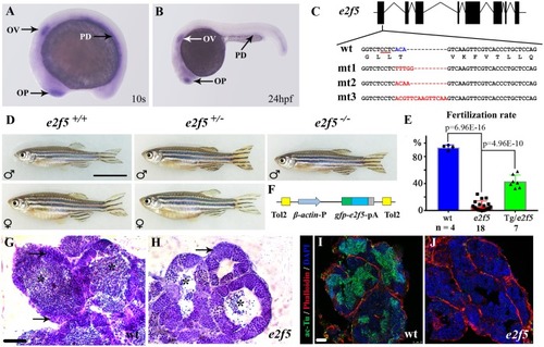
Mutation of <italic>e2f5</italic> leads to male infertility.(A-B) Whole-mount in situ hybridization showing expression of e2f5 at 10-somite stage (10s) and 24 hpf. OP, olfactory placode; OV, otic vesicle; PD, pronephric duct. (C) Genomic structure and sequences of wild- type (wt) and three e2f5 mutant alleles. The underlined sequence in wt indicates PAM sequence of sgRNA target. (D) Phenotypes of male and female wild-type and e2f5 heterozygotes. Only fish exhibiting the male phenotype were present among homozygous mutants. (E) Bar graph showing the percentage of fertilization rates of wild-type, e2f5 homozygote and e2f5 homozygotes carrying Tg(β-actin:gfp-e2f5) transgene as indicated. The numbers of adult males investigated are listed at the bottom. (F) Diagram of the constructs for making gfp-e2f5 transgene. (G-H) H&E staining results showing testes of wild-type (G) and e2f5 homozygous mutants (H). Arrows indicate primary spermatocytes. Asterisks point to mature spermatozoa which were substantially reduced in the mutants. (I-J) Confocal images showing the staining of sperm flagella in wild-type (I) and e2f5 mutant testis (J) visualized with acetylated alpha-tubulin (ac-Tu) antibody. Nuclei and actin filaments were counterstained with DAPI and phalloidin, respectively. Scale bars: 1cm in panel D, 50 μm in panel G, H and 25 μm in panel I, J.
|