FIGURE

Fig. 2

ID
ZDB-FIG-200318-8
Publication
Preston et al., 2019 - A role for OCRL in glomerular function and disease
Other Figures
All Figure Page
Back to All Figure Page
Fig. 2

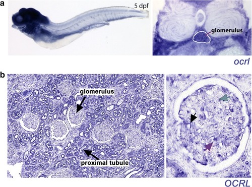
In situ hybridisation detects OCRL expression in the glomeruli of zebrafish and adult human kidney tissue. a Top panels show OCRL expression in a whole-mount zebrafish embryo (left panel, lateral view, anterior to the left) and in transverse section through the glomerulus (right panel, highlighted with white dotted outline) at 5 days post fertilisation (dpf). b In situ hybridisation of normal adult human kidney tissue showing OCRL expression within the glomerulus. The left panel shows a low magnification image with glomeruli and proximal tubules labelled. The right panel shows a close-up view of the glomerulus with OCRL expression labelled in podocytes (black arrow), mesangial cells (green arrow) and endothelial cells (red arrow)

Expression Data
Gene:
Fish:
Anatomical Terms:
Stage: Day 5

Expression Detail
Antibody Labeling
Phenotype Data

Phenotype Detail
Acknowledgments
This image is the copyrighted work of the attributed author or publisher, and ZFIN has permission only to display this image to its users. Additional permissions should be obtained from the applicable author or publisher of the image. Full text @ Pediatr. Nephrol.