|
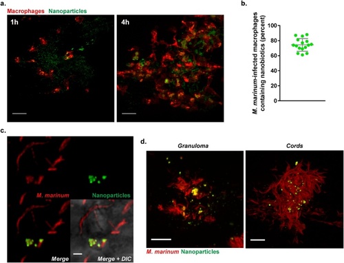
In vivo testing of multi-drug nanobiotics in a M. marinum-infected zebrafish larval model. a. Confocal microscopy images showing nanobiotics-induced macrophage mobilization in vivo. Suspension of coumarin 6-labelled Nano Blank (green) was injected into the muscle of 3 dpf Tg(mpeg1:mCherryCAAX)sh378 zebrafish line harbouring red macrophages. Macrophage chemotaxis towards injection site has been monitored at 1 and 4 h post injection (scale bar, 20 μm). b. Quantification and c. Confocal imaging of coumarin 6-labelled Nano Blank (green) uptake by M. marinum-infected macrophages (red) after 4 h post infection (scale bar, 1 μm). d. Confocal imaging showing the repartition and accumulation of coumarin 6-labelled Nano Blank (green) into a M. marinum (red)-granuloma (left, scale bar, 5 μm) and a mycobacterial cord structure (right, scale bar, 5 μm). (For interpretation of the references to colour in this figure legend, the reader is referred to the web version of this article).
|