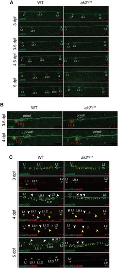
Analysis of lateral line phenotypes using different ak2 mutant transgenic lines. (A) Confocal analysis at different developmental stages of the trunk regions of ak2hg15 embryos and their control siblings (WT) in the Tg(-8.0cldnb:LY-EGFP) background. (B) Confocal analysis from 3.5 to 4 dpf of trunk regions of an ak2hg15 embryo and a control embryo in the Tg(-8.0cldnb:LY-EGFP) background labeling the migrating secondary primordium (dashed ellipse), deposited secondary neuromast (red circle), connecting interneuromast cells and epithelial cells. Confocal images were collected before and right after the end of the time-lapse confocal movie recording. (C) Confocal analysis from 3 to 5 dpf of trunk regions of ak2hg14 embryos and their siblings in different lateral line transgenic backgrounds. Yellow asterisks label the interneuromast cells between L1 and L2 in the sqet20Et transgenic line. White and yellow arrowheads denote primI or primII interneuromast cells, respectively. White asterisks label the migrating secondary primordium. Scale bars: 50 µm.
|

