Figure 4

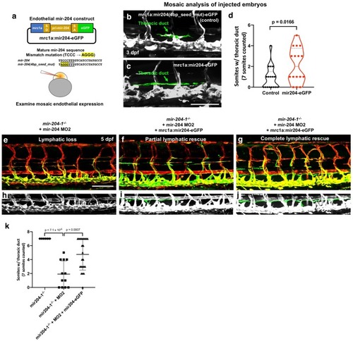
Germline endothelial expression of mir-204 drives precocious lymphatic development.

(a,b) Representative confocal images of mid-trunk of 3 dpf of (a) Tg(mrc1a:eGFP) or (b) Tg(mrc1a:mir204-eGFP). The thoracic duct is pseudocolored in green, with other vessels in gray. (c) Quantification of thoracic duct formation in animals as in panels a and b. The number of somitic segments with an intact thoracic duct was counted, with the same seven mid-trunk somites measured in each animal. (d) Quantification of the area between the dorsal aorta (DA) and posterior cardinal vein (PCV) in animals as in panels a and b, as an indicator of developmental timing. (e) Quantitation of mir-204 level measured by TaqMan qPCR from whole embryo at three dpf. All graphs are analyzed by t-test and the mean ± SD is shown.

Expression Data

Expression Detail
Antibody Labeling
Phenotype Data

Phenotype Detail
Acknowledgments
This image is the copyrighted work of the attributed author or publisher, and ZFIN has permission only to display this image to its users. Additional permissions should be obtained from the applicable author or publisher of the image. Full text @ Elife