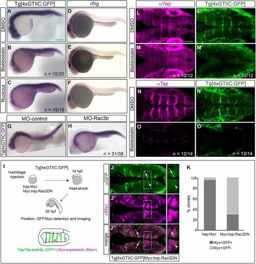
Yap and Taz in the hindbrain boundaries sense mechanical cues. (A-C,G,H) Whole-mount gfp in situ hybridization of Tg[4×GTIIC:GFP] embryos treated with DMSO (A), with myosin II pharmacological inhibitors such as blebbistatin (B) and rockout (C) from 16 to 22 hpf, or injected with MO-control (G) or MO-Rac3b (H) in order to downregulate Rac3b. In all distinct experimental cases, gfp expression, and therefore Yap/Taz activity, is downregulated in the hindbrain boundaries and is not affected in the somites. (D-F) Whole-mount rfng in situhybridization of Tg[4xGTIIC:GFP] embryos treated with DMSO (D), blebbistatin (E) or rockout (F). Expression of boundary markers is not affected upon treatment, as previously shown (Gutzman and Sive, 2010). Lateral views with anterior to the left. (I-K) Downregulation of Rac3b by clonal analysis. (I) Scheme depicting the functional experiment in which Tg[4xGTIIC:GFP] embryos were injected at the one-cell stage with inducible Myc-tagged constructs (hsp:Myc or Myc:hsp:Rac3DN), heat-shocked at 14 hpf, allowed to develop until 30 hpf, and immunostained for GFP and Myc. For the phenotypic analysis, we scored the percentage of Myc-expressing clones (Myc-positive) hitting the boundaries that displayed Yap/Taz-TEAD activity (GFP-positive) and this was plotted in K. (J-J″) Example of a Tg[4xGTIIC:GFP] embryo injected with Myc:hsp:Rac3DN and immunostained using anti-GFP (green) and anti-Myc (magenta) antibodies. Myc-positive cells, when located within the boundaries, display low (see arrows) or no (see arrowheads) GFP-expression. On the right are images from the regions framed in J-J″ showing a magnified boundary in which the Yap/Taz-TEAD activity has been either completely abolished or downregulated upon expression of Rac3DN. Dorsal view with anterior to the left. (K) The percentage of boundary cell clones expressing Yap/Taz-TEAD activity (GFP-positive clones) are displayed in dark gray in the histogram, over the total Myc-positive clones displayed as light gray in the histogram, either in control (hsp:Myc) or experimental conditions (Myc:hsp:Rac3DN) where the actomyosin cables were compromised. When Rac3b is downregulated, the percentage of Myc-positive clones with Yap/Taz-TEAD activity decreases. (L-O′) Tg[4xGTIIC:GFP] embryos treated with DMSO (L,L′,N,N′) or with blebbistatin (M,M′,O,O′) from 19-25 hpf, and immunostained using anti-Yap (L,M) or anti-Taz (N,O) antibodies, and analyzed for TEAD activity (L′,M′,N′,O′). Expression of GFP and Yap and Taz in the boundary cells is abolished upon blebbistatin treatment. Dorsal views with anterior to the left. r, rhombomere. n=X/Y indicates the number of embryos with the displayed phenotype (X) over the total number of analyzed embryos (Y). Scale bars: 200μm in A-H; 50μm in J-J″,L-O.

