
Fig. S5
- ID
- ZDB-FIG-190820-40
- Publication
- Zhu et al., 2019 - Aplnra/b Sequentially Regulate Organ Left-Right Patterning via Distinct Mechanisms
- Other Figures
- All Figure Page
- Back to All Figure Page
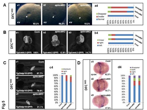
aplnra or apela loss of function in DFCs resulted in LR patterning defect. (A. a1-a4) The KV phenotype in control and aplnra or apela loss of function in DFCs. At 10 somite stage, 56.3% and 10.2% of embryos injected with aplnra MO in DFCs displayed smaller KV and disappeared KV(A. a2-a4, n=119), 57.6% and 12.0% of embryos injected with apela MO in DFCs displayed smaller KV and disappeared KV (A. a4, n=90), while 92.2% of control embryos displayed normal KV (A. a1 and a4, n=103). (B. b1-b4) At 60hpf, part of embryos injected with aplnra MO displayed linear heart (B. b2 and b4, 14.7%, n=116) and reversed heart (B. b3 and b4, 12.9%, n=116), 15.5% and 13.1% of embryos injected with apela MODFCs displayed reversed heart and linear heart (B. b4, n=90). (C. c1-c3) Liver LR defect was also observed in aplnra MO (total 21.1%, n=95) or apela MO (total 24.8. %, n=90) injected embryos in DFCs (C. c3 and c4). (D. d1-d4) spaw expression was examined in embryos, right-sided and both-sided spaw was discovered in 17.9% and 10.7% of embryos with aplnra loss of function in DFCs (D. d2-d4, n=56) or 15.5%, 11.1% and 8.9% of embryos displayed right-sided, both-sided and disappeared spaw with apela loss of function in DFCs(Dd4, n=56) |