
Fig. 3
- ID
- ZDB-FIG-190816-11
- Publication
- Kuroda et al., 2018 - Roles of basal keratinocytes in actinotrichia formation
- Other Figures
- All Figure Page
- Back to All Figure Page
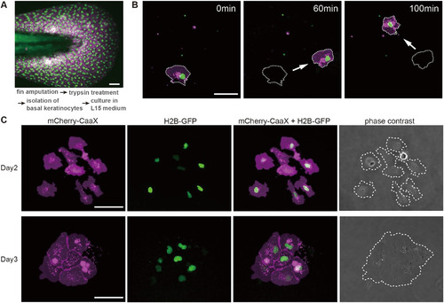
Primary culture of the basal keratinocytes from the reporter zebrafish expressing fluorescent proteins at larval stage. (A) Median fin fold of larva at 3 days post-fertilizations (dpf) expressing histone H2B-GFP which can visualize the nucleus and mCherry-CaaX in the basal keratinocytes. Basal keratinocytes were isolated from the median fin fold by the trypsin treatment and cultured in the L15 medium. (B) Time-lapse images of basal keratinocytes. White arrow indicates the direction of basal keratinocyte movement. (C) Basal keratinocytes can self-assemble and form colonies. The images on the top row show 8 basal keratinocytes which were separated one another after 2 days in culture. The images on the bottom row show a colony that consists of 8 basal keratinocytes at 3 days after culture. White dot lines show the shape of a cell or a colony. Scale bar is 50 μm (A–C). |
Reprinted from Mechanisms of Development, 153, Kuroda, J., Iwane, A.H., Kondo, S., Roles of basal keratinocytes in actinotrichia formation, 54-63, Copyright (2018) with permission from Elsevier. Full text @ Mech. Dev.