|
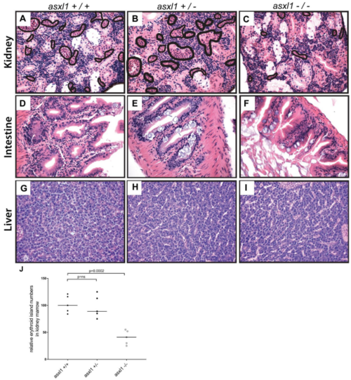
Histopathological analysis of kidney, intestine and liver development in 17-month-old asxl1+/+ and asxl1+/- and asxl1-/- fish. Results revealed that intestine (D, E and F) and liver (G, H and I) was normal and similar to that of the fish, indicating that these fish recovered from early developmental hypoplasia in these organs. However, asxl1-/- mutants did have decreased numbers of erythroid islands in the kidney marrow compared with asxl1+/+ and asxl1+/- fish (A, B and C), indicating that the hematopoietic system is abnormal in these animals. Area encircled with black line indicates erythroid islands. The quantification of erythroid islands was shown in (J), with the black bars representing the median values. ns p>0.05, *p<0.05, **p<0.01 by two-tailed unpaired t-test.
|

