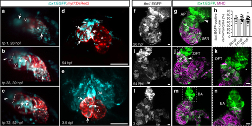
tbx1+ myocardial precursors connect to the FHF myocardium during heart tube stages. a–c Maximum intensity projections of representative stages of a high-resolution reconstruction of the beating heart of a tbx1:EGFP;myl7:DsRed2 double-positive transgenic between 28 and 52 hpf; lateral view (right side) of the embryo, anterior to the top, ventricle to the upper left, atrium to the lower right, and cardiac imaging phase 27 (n = 1); ventricle (V), atrium (A), and bulbus arteriosus (BA). Arrows indicate tbx1+/myl7- cells at the OFT and IFT at the beginning of the time-lapse (a) that gradually turn on myl7 reporter expression (b, c). The dashed line (b, c) indicates the distal end of the ventricle and the arrowheads point to tbx1:EGFP-expressing cells at the OFT that never cross into the ventricle and are likely BA precursors. d, e Maximum intensity projections of SPIM-imaged tbx1:EGFP;myl7:DsRed2 double-positive transgenic hearts stopped from contracting with BDM at 54 hpf (n = 3) or 3.5 dpf (n = 7), respectively; ventral views, anterior to the top. tbx1 reporter expression can be detected in differentiated tbx1/myl7 reporter double-positive cardiomyocytes (arrows d, e), the tbx1+/myl7- OFT at 54 hpf (arrowhead d), and the tbx1+/myl7- BA at 3.5 dpf (asterisk e). f, g, i–n Top-down 2-µm confocal section of isolated zebrafish hearts at 26 (f, g), 54 (i–k), and 72 hpf (l–n) from tbx1:EGFP, counterstained with anti-GFP and anti-MHC; OFT/BA to the top, sinoatrial node (SAN) or atrium (A) to the bottom left, and ventricle (V) to the top left. k, n A magnification of the framed area in j, m. f, gtbx1:EGFP is expressed at the MHC-negative arterial pole (AP) of the heart tube (arrowhead). h Quantification of the tbx1:EGFP-positive ventricular cardiomyocytes compared to whole ventricle (in percentage) reveals no change over developmental period from 26 hpf (n = 5), 54 hpf (n = 11), to 72 hpf (n = 15). Each data point represents averaged percentage per heart; means ± s.d. i–k At 54 hpf, cardiomyocytes of the later-differentiated distal ventricle express tbx1:EGFP (arrowhead), as do MHC-negative progenitors of the OFT (asterisks). l–n The differentiated BA at 3 dpf is positive for tbx1:EGFP. Scale bars 200 µm (d, e), 10 µm (f, g, i–n)
|
