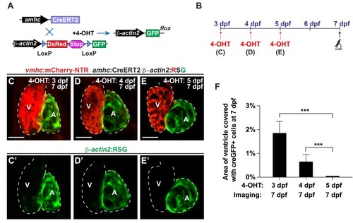
Figure 1—figure supplement 2.

Reprogramming of atrial cardiomyocytes into ventricular cardiomyocytes during development.

(A) Schematic representation of the transgenic lines utilized in lineage tracing experiments. Treatment with 4-hydroxytamoxifen (4-OHT) activates the amhc:CreERT2 protein, which causes the recombination of β-actin2:RSG into β-actin2:GFP, thus permanently labeling amhc expressing cells in green. (B) vmhc:mCherry-NTR; amhc:CreERT2; β-actin2:RSG hearts were exposed to tamoxifen at (C) 3, (D) 4 or (E) 5 dpf in order to genetically label atrial CMs with GFP (c-aGFP), and then confocal imaged at 7 dpf. These hearts that were genetically labeled at (C) 3 (n = 21), (D) 4 (n = 14) or (E) 5 (n = 22) dpf show varying contributions of c-aGFP+ CMs to the ventricles. Green channel – (C’–E’) genetically labeled c-aGFP+ atrial CMs. (F) Quantitation of the percentage of ventricular area covered with c-aGFP+ CMs reveals that although uninjured hearts genetically labeled at 3 and 4 dpf exhibit a small contribution of c-aGFP+ CMs to the ventricle, uninjured hearts labeled at 5 dpf do not. All confocal images shown are maximum intensity projections. V, ventricle; A, atrium; dpf, days post-fertilization. Dashed lines outline the heart. Bars: 50 μm. Mean + s.d. ANOVA test, ***, p<0.001.

Expression Data

Expression Detail
Antibody Labeling
Phenotype Data

Phenotype Detail
Acknowledgments
This image is the copyrighted work of the attributed author or publisher, and ZFIN has permission only to display this image to its users. Additional permissions should be obtained from the applicable author or publisher of the image. Full text @ Elife