
Fig. 2
- ID
- ZDB-FIG-190627-13
- Publication
- Jung et al., 2019 - Znf76 is associated with development of the eyes, midbrain, MHB, and hindbrain in zebrafish embryos
- Other Figures
- All Figure Page
- Back to All Figure Page
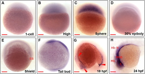
Spatiotemporal expression of zebrafish znf76 during embryonic development WISH experiments performed with a znf76 antisense RNA probe at the 1-cell (A), high (B), sphere (C), 30% epiboly (D), shield (E), tail bud (F), 18 hpf (G), and 24 hpf (H) stages. All the images are from the lateral view (A–H). znf76transcripts were zygotically expressed in the early stages and were reduced in the 30% epiboly and shield stages (D,E). znf76 transcripts were present in the head and tail region (red arrowheads) at the tail bud stage (F) and then became progressively restricted to the trigeminal placode and proctodeum posterior gut, with a significant reduction in expression by the late somite stage (18 hpf) (G). At 24 hpf (H), znf76 transcripts were restricted to Mb, MHB, and Hb-N. Red arrowheads show the anatomical structures. Mb – midbrain, MHB – midbrain–hindbrain boundary, Hb-N – hindbrain neurons, E – eye, ES – Embryonic shield. Scale bar: 100 µm |
| Gene: | |
|---|---|
| Fish: | |
| Anatomical Terms: | |
| Stage Range: | 1-cell to Prim-5 |