
Fig. S4
- ID
- ZDB-FIG-190620-29
- Publication
- Ofer et al., 2019 - A novel nonosteocytic regulatory mechanism of bone modeling
- Other Figures
- (all 9)
- All Figure Page
- Back to All Figure Page
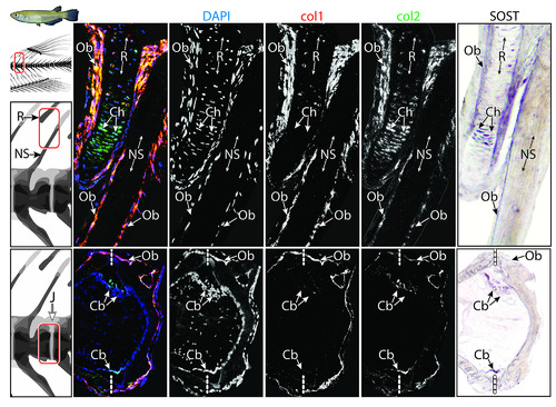
Top row: NS and adjacent fin R; bottom row: intervertebral J. Each row comprises (from left to right) a multichannel RGB double fluorescent ISH image, followed by isolated single-channel images (DAPI, col1, col2, respectively) and a SOST ISH image. The figure provides a deconstruction of the multichannel ISH images Fig 4. Note SOST-positive Cbs, Chs, and Obs and the lack of osteocytes within the NS. Refer to text for further results. Cb, chordoblast; Ch, chondrocyte; ISH, in situ hybridization; J, joint; NS, neural spine; Ob, osteoblast; R, radial; RGB, red-green-blue. |