
Fig. 6
- ID
- ZDB-FIG-190530-40
- Publication
- Wong et al., 2018 - ERK Activity Dynamics during Zebrafish Embryonic Development
- Other Figures
- All Figure Page
- Back to All Figure Page
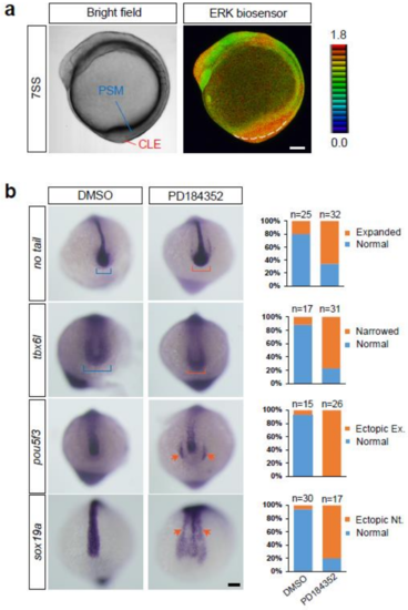
Roles of ERK activity in the tail bud. (a) Bright field (left panel) and ERK activity (right panel) of the embryo at the 7-somite stage. PSM, pre-somitic mesoderm; CLE, caudal lateral epiblast. Lateral view, anterior to the top. Scale bar, 100 m. (b) Expression of no tail, tbx6l, pou5f3, and sox19a in embryos treated with DMSO (vehicle) or the MEK inhibitor PD184352. In MEK inhibitor-treated embryos, a pool of axial stem cells (caudal region of the neural tube), which is marked by no tail expression, expanded (orange bracket, the first row from the top), and the paraxial mesoderm, marked by tbx6l, became narrower (orange bracket, second row from the top). In addition, ectopic neural tube formation, as evidenced by expression of pou5f3 (a marker of neural progenitors; orange arrows, third row from the top) and sox19a (orange arrows, fourth row from the top), was frequently induced in MEK inhibitor-treated embryos. Scale bar, 100 m. |