
Fig. 5
- ID
- ZDB-FIG-180921-29
- Publication
- Gistelinck et al., 2018 - Zebrafish type I collagen mutants faithfully recapitulate human type I collagenopathies
- Other Figures
- All Figure Page
- Back to All Figure Page
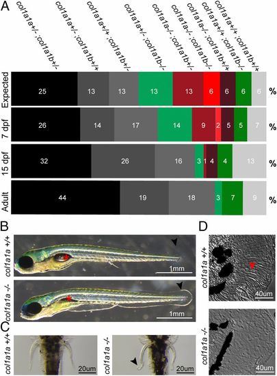
In-depth analysis of mutants with partial or complete loss of col1a1a and/or col1a1b. (A) Segregation analysis of the different mutant genotypes generated from an in-cross of col1a1a+/−;col1a1b+/− double-heterozygous mutant fish. Progeny were genotyped at three different time points (100 fish per time point): 7 dpf, 15 dpf, and adult stage (3 mo). The relative abundance of each genotype is given for each time point as a percentage. “Expected” indicates the theoretical abundance of each genotype, following Mendelian segregation. All combinations with a complete loss of col1a1a (col1a1a−/−, indicated in red shades), are less viable and completely lost when progeny reached adulthood. Genotypes that contain a complete loss of col1a1b (col1a1b−/−, indicated in green shades) are still present at adult stage. Black indicates the parental genotype, white indicates wild-type fish, and gray represents the other remaining genotypes. (B) Phenotype of col1a1a−/− mutant larvae at 7 dpf, compared with wild-type siblings. Col1a1a−/− mutant larvae present with a noninflated swimbladder (asterisk) and a ruffled finfold (arrowhead). (C) Ventral view and detail of the pectoral fins showing frilly distal margins (arrowhead) in the pectoral fins of col1a1a−/− mutant larvae at 7 dpf. (D) Detail of the col1a1a−/− mutant larvae finfold at 7 dpf, showing an absence of fin actinotrichia fibers, which can be clearly observed in the finfold of wild-type siblings (arrow). |
| Fish: | |
|---|---|
| Observed In: | |
| Stage: | Days 7-13 |