
FIGURE
Fig. S4
Fig. S4
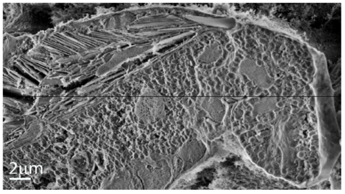
Cryo-SEM image of the zebrafish iris at 21 dpf. Image of a freeze-fractured iris shows the area adjacent to the lens, where two layers of iridophores are visible. Scale bar: 2 μm. Figure |
Expression Data
Expression Detail
Antibody Labeling
Phenotype Data
Phenotype Detail
Acknowledgments
This image is the copyrighted work of the attributed author or publisher, and
ZFIN has permission only to display this image to its users.
Additional permissions should be obtained from the applicable author or publisher of the image.
Full text @ Adv Sci (Weinh)