Fig. 7
- ID
- ZDB-FIG-180608-51
- Publication
- Sun et al., 2018 - Transcripts within rod photoreceptors of the Zebrafish retina
- Other Figures
- All Figure Page
- Back to All Figure Page
|
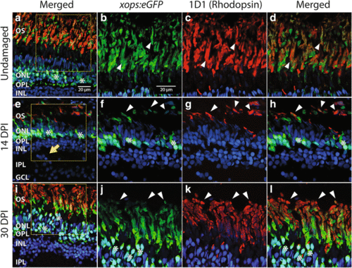
Regenerated rods in xops:eGFP transgenic zebrafish. a Undamaged retina, showing GFP+ (green); rod cell bodies in the outer nuclear layer (ONL) and GFP+, rhodopsin (1D1, magenta) outer segments (OS). Section is counterstained with DAPI (nuclei; blue). Asterisks (*) highlight some of the GFP+, DAPI+ cell bodies in the outer nuclear layer (ONL). Boxed area is shown at higher magnification in (b-d). b-d. Higher magnification views of outer segments of GFP imaging channel only (b), rhodopsin only (c), and a merged image (d), to show colabeling of outer segments (OS; some are highlighted by arrowheads). e-h Regenerated retina processed 14 days after injury (14 dpi), low magnification view with GPF, rhodopsin, and DAPI (e), and higher magnification views of GFP (f), rhodopsin (g), and merged image (h), to show colabeling of outer segments in regenerated retina (some are highlighted by arrowheads). Arrow in e shows example of histological error, with a cluster of DAPI+ cell bodies misplaced in the inner plexiform layer (IPL). i-l Regenerated retina processed 30 days after injury (30 dpi), low magnification view with GPF, rhodopsin, and DAPI (i), and higher magnification views of GFP (j), rhodopsin (k), and merged image (l), to show colabeling of outer segments in regenerated retina (some are highlighted by arrowheads). Boxed area in (i) is shown at higher magnification in (j-l). In b-d, f-h, and j-k, note that GFP fluorescence can be brighter than the corresponding 1D1 fluorescence, or vice-versa, although all OS show colabeling. Scale bar in a (applies to a, e, i) = 20 μm. Scale bar in b (applies to b-d, f-h, j-l) = 20 μm. OPL, outer plexiform layer; INL, inner nuclear layer; GCL, ganglion cell layer |
| Genes: | |
|---|---|
| Antibody: | |
| Fish: | |
| Condition: | |
| Anatomical Terms: | |
| Stage: | Adult |

