
Fig. 1
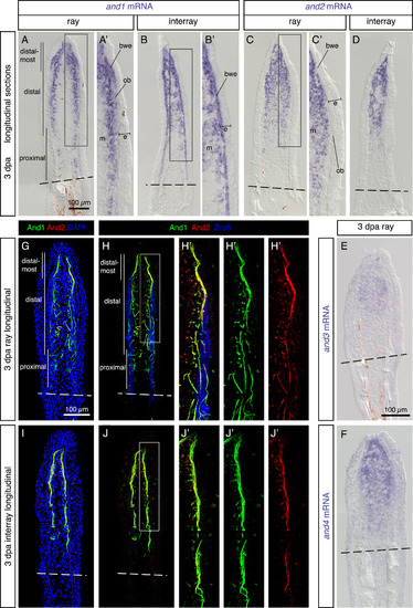
Analysis of the actinotrichia-specific components in the regenerative outgrowth. (A-F) In-situ hybridization with probes against four actinodin genes on longitudinal fin sections at 3 dpa. N ≥ 4 fins; ≥ 3 sections per fin. (A, B) and1 mRNA is detected in the mesenchyme (m) of the blastema and in the basal wound epithelium (bwe). No expression is detected in the basal wound epithelium adjacent to osteoblasts (ob) in the ray. (C, D) and2 (E) and3 (F) and4 genes are co-expressed in the mesenchyme of the blastema, but not in the epidermis (e). (G-J) Immunofluorescence staining of longitudinal fin sections of ray (G, H) and interray (I, J) at 3 dpa. And1 (green) and And2 (red) proteins are co-localized in actinotrichia fibers. Rays and interrays are identified by the presence versus absence of Zns5-positive osteoblasts (H, J, blue). Actinotrichia fibers are predominantly found in the subepithelial position with the exception of osteoblast-containing regions. N = 4 fins; 3 sections per fin. Higher magnifications of framed areas are labeled with the same letter with a prime symbol. Fin amputation planes are shown with a dashed line. The same rules apply to all subsequent figures. |
| Genes: | |
|---|---|
| Antibodies: | |
| Fish: | |
| Condition: | |
| Anatomical Terms: | |
| Stage: | Adult |
Reprinted from Developmental Biology, 433(2), König, D., Page, L., Chassot, B., Jaźwińska, A., Dynamics of actinotrichia regeneration in the adult zebrafish fin, 416-432, Copyright (2017) with permission from Elsevier. Full text @ Dev. Biol.