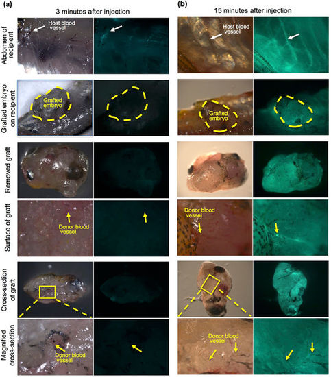
Fig. 2
- ID
- ZDB-FIG-180420-11
- Publication
- Kawasaki et al., 2017 - Development and growth of organs in living whole embryo and larval grafts in zebrafish
- Other Figures
- All Figure Page
- Back to All Figure Page
|
Microinjection of FITC-dextran into the recipient heart. To examine vascularization between recipients and grafted embryos, FITC-conjugated dextran solution was injected into the hearts of living recipients in which 72 hpf embryos had been transplanted 3 months previously. (a) At 3 minutes after injection, slight fluorescence of the recipient blood vessels (white arrow) was observed in the recipient abdomen but not in grafted embryos (yellow dotted line). No fluorescence in the blood vessels was observed in the grafted embryo (yellow arrows). The recipient heart in which FITC-dextran was injected is outside the frame of this panel. Most of the FITC-dextran has not reached the abdomen at 3 minutes after injection. (b) At 15 minutes after injection, strong fluorescence was observed in the recipient blood vessels (white arrow) of the abdomen and the grafted embryos (yellow dotted line). This strong fluorescence was also observed in the blood vessels of the grafted embryo (yellow arrows). These results suggest that recipient blood vessels are connected with grafted embryos. |

