|
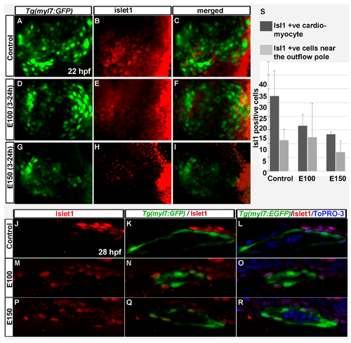
Embryonic ethanol exposure reduced second heart field precursors. (A–I) Anti-Islet1 antibody stained Tg(myl7:GFP) embryos showed Islet1 positive second heart field precursors in the rotating heart cone in control embryos (A–C) at 22 hpf; ethanol exposed embryos showed reduced number of Islet1 positive cells in the heart cones (D–I) at 22 hpf; (J–R) Anti-Islet1 antibody stained Tg(myl7:GFP) embryos showed second heart field derived cardiomyoctes in the heart (Myl7 and Islet1 double positive; red and green) and second heart field precursors (Islet1 positive, red) near the out flow pole in the control embryos (J–L) and ethanol-exposed embryos (M–R) at 28 hpf; (S) Graph shows the quantification of the Islet1/Myl7 double positive cardiomyocytes in the heart tube and Islet1 positive cells near the outflow pole at 28 hpf.
|