Fig. 3
- ID
- ZDB-FIG-180326-3
- Publication
- Watanabe et al., 2017 - Coordinated Expression of Two Types of Low-Threshold K+ Channels Establishes Unique Single Spiking of Mauthner Cells among Segmentally Homologous Neurons in the Zebrafish Hindbrain.
- Other Figures
- All Figure Page
- Back to All Figure Page
|
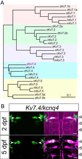
Expression of Kv7.4/Kcnq4 mRNA in M cells during development. A, Phylogenetic tree of α-subunit proteins of XE991-sensitive voltage-gated K+ channel Kv7/KCNQ family members in zebrafish, Xenopus, chick, mouse, and human (z, x, c, m, and h as the gene prefix, respectively), and showing identification of zebrafish Kv7.4 (magenta). B, Dorsal views of rhombomeres 4, 5, and 6 (r4, r5, and r6) in the hindbrain at 2 dpf (top) and 5 dpf (bottom) after in situ hybridization using a Kv7.4 antisense probe (green). Kv7.4 mRNA was expressed in M cells (arrowhead) at 2 and 5 dpf, but not in MiD2cm and MiD3cm cells, labeled (magenta) immunohistochemically (with 3A10 antibody, at 2 dpf) or retrogradely (at 5 dpf). Scale bar, 20 μm. |
| Gene: | |
|---|---|
| Antibody: | |
| Fish: | |
| Anatomical Terms: | |
| Stage Range: | Long-pec to Day 5 |

