
Fig. 4
- ID
- ZDB-FIG-180117-29
- Publication
- Nam et al., 2017 - Harmless effects of argon plasma on caudal fin regeneration and embryogenesis of zebrafish: novel biological approaches for safe medical applications of bioplasma
- Other Figures
- All Figure Page
- Back to All Figure Page
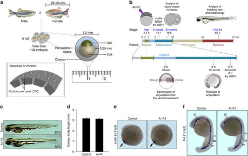
Biosafety of Ar-PJ with respect to the biogenesis of living organisms—experimental validations of embryogenesis as a novel biosafety assessment system. (a) Schematic diagrams of zebrafish fertilization and the structure of an embryo at 0 hpf. (b) A schematic diagram of experimental designs for assessing zebrafish embryogenesis. (c) Evaluation of the completion of embryogenesis in response to Ar-PJ, a visual indicator of Ar-PJ biosafety. Hatched embryos were counted at 60 hpf, and the hatching rate was determined by calculating the hatching percentage (H%) for each sample: H%=(the number of hatched embryos/the total number of embryos) × 100. Lateral view, anterior to the top. (d) Quantification of body length—a measurable indicator of Ar-PJ biosafety assessment. The lateral length of embryos at 60 hpf was measured, and values are presented as the means±s.e.m. (control n=7, Ar-PJ n=12). (e) Evaluation of Ar-PJ biosafety at the molecular level. Live zebrafish embryos at the blastula stage (3.3 hpf) were treated for 30 s with Ar-PJ. WISH was performed with a wnt8a antisense probe on segmentation-period embryos (12 hpf, 5-somite stage). (f) Evaluation of blood vessel morphogenesis, an embryonic visual indicator for Ar-PJ biosafety assessment. WISH was performed with a fli antisense probe on segmentation-period embryos (19 hpf). Vasculogenesis sites are indicated: pharyngeal (pr), dorsal aorta (da), axial vein (av), intersegmental vessels (iv) and intermediate cell mass (icm). |