|
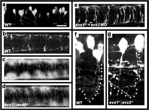
V0v cells develop into CoSA interneurons. Immunohistochemistry for EGFP in Tg(evx1:EGFP) SU1 (a, b & e) or Tg(evx1:EGFP) SU2 (c, d, f & g) embryos. . a, f & g lateral views with dorsal up and anterior left of spinal cord at 27 h (a) or 48 h (f & g). b-e dorsal views with anterior left of zebrafish spinal cord at 30 h (b) or 48 h (c-e). a-c & f WT, (d & g) evx1;evx2 double mutant, (e) evx1 mutant injected with evx2 morpholino. b & c show increasing number of commissural axons crossing the spinal cord as development proceeds. d & e demonstrate that V0v axons are still clearly commissural in the absence of Evx1 and Evx2. f & g show magnified views of commissural ascending V0v axons. White arrows (drawn slightly to the right of the axon so that EGFP expression is still visible) indicate ascending axon trajectories. Scale bar: 50 μm (a-e) and 15 μm (f & g)
|