
FIGURE
Fig. 7
- ID
- ZDB-FIG-171002-13
- Publication
- Fernández et al., 2017 - Optimizing pulse compressibility in completely all-fibered Ytterbium chirped pulse amplifiers for in vivo two photon laser scanning microscopy
- Other Figures
- All Figure Page
- Back to All Figure Page
Fig. 7
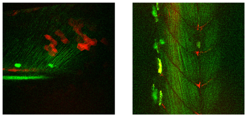
Image of a 5 days old zebrafish larva’s tail (left) and 2 days old larva’s muscle tissue (right). The FOV of both images is 300x300 µm2, the images are composed of 100 and 77 frames respectively. The green line structures are resulting from second harmonic radiation generated from collagen fibrils. The red structures are due to two-photon fluorescence from mCherry labelled cells. The (large) bright green/yellow structures visible are pigmented cells with stronger scattering. |
Expression Data
Expression Detail
Antibody Labeling
Phenotype Data
Phenotype Detail
Acknowledgments
This image is the copyrighted work of the attributed author or publisher, and
ZFIN has permission only to display this image to its users.
Additional permissions should be obtained from the applicable author or publisher of the image.
Full text @ Biomed. Opt. Express