
Fig. S1
- ID
- ZDB-FIG-170922-54
- Publication
- Xue et al., 2017 - The Vascular Niche Regulates Hematopoietic Stem and Progenitor Cell Lodgment and Expansion via klf6a-ccl25b
- Other Figures
- All Figure Page
- Back to All Figure Page
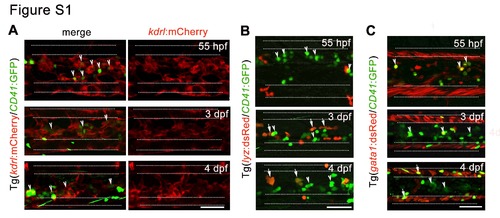
Figure S1. The anatomical relationship between HSPCs and perivascular niche cells. Related to Figure 1. (A) Confocal imaging of Tg (kdrl:mCherry/CD41:GFP) CHT region showing the localization of HSPCs adjacent to vascular ECs. (B-C) Confocal imaging in different transgenic embryos displaying the location of CD41+ cells and hematopoietic cells, including lyz:dsRed+ myeloid cells (B) and gata1:dsRed+ erythroid cells (C) in the CHT. The arrowheads indicate HSPCs and arrows mark differentiated cells derived from HSPCs. The white dashed lines mark the outlines of caudal artery and caudal vein respectively. Scale bar, 50 μm. |
Reprinted from Developmental Cell, 42(4), Xue, Y., Lv, J., Zhang, C., Wang, L., Ma, D., Liu, F., The Vascular Niche Regulates Hematopoietic Stem and Progenitor Cell Lodgment and Expansion via klf6a-ccl25b, 349-362.e4, Copyright (2017) with permission from Elsevier. Full text @ Dev. Cell