
FIGURE
Fig. S10
- ID
- ZDB-FIG-170921-50
- Publication
- Kara et al., 2017 - miR-27 regulates chondrogenesis by suppressing Focal Adhesion Kinase during pharyngeal arch development
- Other Figures
- All Figure Page
- Back to All Figure Page
Fig. S10
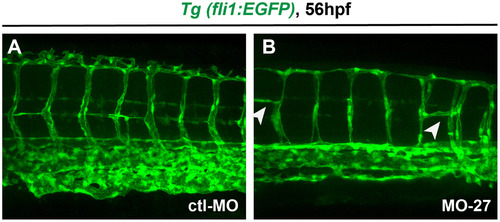
Vasculature defects in miR-27 morphants. Vasculature patterning in live Tg(fli1a:eGFP)y1 embryos at 56 hpf. Embryos were injected with either MO-ctl or MO-27 at the single cell stage. Arrowheads indicate branching defects in the intersegmental vessels in miR-27 morphants. |
Expression Data
Expression Detail
Antibody Labeling
Phenotype Data
Phenotype Detail
Acknowledgments
This image is the copyrighted work of the attributed author or publisher, and
ZFIN has permission only to display this image to its users.
Additional permissions should be obtained from the applicable author or publisher of the image.
Reprinted from Developmental Biology, 429(1), Kara, N., Wei, C., Commanday, A.C., Patton, J.G., miR-27 regulates chondrogenesis by suppressing Focal Adhesion Kinase during pharyngeal arch development, 321-334, Copyright (2017) with permission from Elsevier. Full text @ Dev. Biol.