
Fig. S1
- ID
- ZDB-FIG-170914-48
- Publication
- Tornini et al., 2017 - Live fate-mapping of joint-associated fibroblasts visualizes expansion of cell contributions during zebrafish fin regeneration
- Other Figures
- All Figure Page
- Back to All Figure Page
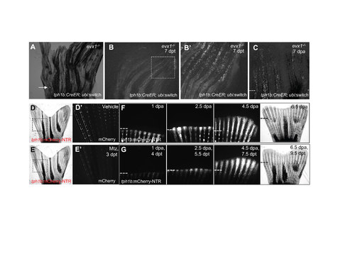
Joint-associated tph1b+ cell progeny are dispensable for fin regeneration. (A) tph1b:CreER; ubi:switch; evx1-/- fish were treated with 4 μM tamoxifen to induce recombination and cell labeling. A few cells in crushed areas (arrow) were labeled. (B) tph1b:CreER; ubi:switch; evx1-/- fish were treated with 4 μM tamoxifen to induce recombination and cell labeling. Rays with more severe injuries show labeled tph1b+ cells at 7 days post-treatment (dpt). Box in (B) magnified in (B’). (C) tph1b:CreER; ubi:switch; evx1-/- fish were treated with 4 μM tamoxifen, and rays with labeled cells were amputated at 7 dpt and tracked through 7 dpa. Injury-induced tph1b+ cells can contribute to regenerating fin tissue. Dashed line, amputation plane. (D, E) tph1b:mCherry-NTR fish were treated with vehicle (D) or metronidazole (Mtz) (E). mCherry signal for boxes in (D) and (E) are shown in (D’) and (E’) at 3 days posttreatment (dpt). (F, G) Fins of vehicle (F)- or Mtz (G) -treated tph1b:mCherry-NTR fins were amputated and imaged at at 1, 2.5, 4.5, or 6.5 dpa. No gross differences in fin regeneration were observed following tph1b+ cell ablation. n = 6 for vehicle-treated; n = 4 for Mtz-treated. Dashes lines, amputation plane. |
| Gene: | |
|---|---|
| Fish: | |
| Conditions: | |
| Anatomical Terms: | |
| Stage: | Adult |