
FIGURE
Fig. S1
- ID
- ZDB-FIG-170524-20
- Publication
- Albuixech-Crespo et al., 2017 - Molecular regionalization of the developing amphioxus neural tube challenges major partitions of the vertebrate brain
- Other Figures
- All Figure Page
- Back to All Figure Page
Fig. S1
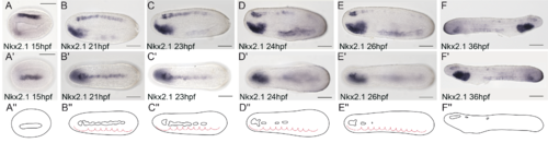
Temporal expression of Nk2.1 during amphioxus development. Lateral views (A-F, F’), dorsal views (A’-E’), and schematic drawings (A''-F'') of the neural component of Nk2.1 gene expression pattern from 15 to 36 hours post-fertilization. Anterior is to the left except in F. Somites are indicated using red dotted lines. Scale bar = 50μm. |
Expression Data
Expression Detail
Antibody Labeling
Phenotype Data
Phenotype Detail
Acknowledgments
This image is the copyrighted work of the attributed author or publisher, and
ZFIN has permission only to display this image to its users.
Additional permissions should be obtained from the applicable author or publisher of the image.
Full text @ PLoS Biol.