
Fig. S4
- ID
- ZDB-FIG-170516-31
- Publication
- Münch et al., 2017 - Notch signalling restricts inflammation and serpine1 expression in the dynamic endocardium of the regenerating zebrafish heart.
- Other Figures
- All Figure Page
- Back to All Figure Page
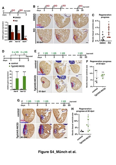
Notch signalling modulation impairs heart regeneration (A) Relative gene expression (qPCR) of the Notch target genes dll4, her15, and her4, showing reduced expression after RO treatment (indicated on top). * P<0.05; **P<0.01; ***P<0.005. (B) Representative AFOG-stained sections from three levels of the hearts of fish treated with DMSO or RO for 30 dpci (treatment regime is indicated on top). Cardiac muscle: brown; fibrin: pink/ orange; collagen: blue. (C) Scatter plot showing the amount of fibrotic tissue relative to ventricle size in hearts from fish treated with DMSO or RO (discontinuous line = mean ± s.d, t-test *P<0.05) (D) qPCR showing higher expression of her4 and her15 in injured hearts of transgenic Tg(UAS:NICD) fish compared than in the control at 3 dpci. (heat shock regime is indicated on top ( mean ± s.d, t-test, ***P< 0.005). (E) AFOG–stained sections taken at 3 anatomical levels of WT and Tg(UAS:NICD) hearts at 33 dpci, showing similar regenerative capacity (heat shock regime is indicated on top). (F) Quantification of the progress of regeneration at 33 dpci (discontinuous line = mean± s.d, t-test, not significant). (G) AFOG-stained sections taken at 3 anatomical levels of WT and Tg(UAS:NICD) hearts at 90 dpci, showing failed regeneration (heat shock regime is indicated on top). (H) Quantification of the progress of regeneration at 90 dpci (discontinuous line = mean, t-test *P<0.05). Scale bars: 100 μm. |
| Fish: | |
|---|---|
| Conditions: | |
| Observed In: | |
| Stage: | Adult |