
FIGURE
Fig. S2
- ID
- ZDB-FIG-170209-44
- Publication
- Noack Watt et al., 2016 - The Roles of RNA Polymerase I and III Subunits Polr1c and Polr1d in Craniofacial Development and in Zebrafish Models of Treacher Collins Syndrome
- Other Figures
- All Figure Page
- Back to All Figure Page
Fig. S2
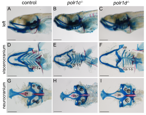
polr1c and polr1d mutant embryos have craniofacial cartilage anomalies at 9 dpf. (A-C) Alcian blue and Alizarin red staining reveals diminished cartilage and bone formation in polr1c and polr1d mutant embryos. (D-F) Dissection of the viscerocranium revealed mispatterning of the ceratohyal (ch), and hypoplasia of the ceratobranchial cartilages (cb) in polr1c and polr1d mutants. There is also hypoplasia of bone elements including the opercles (op) and pharyngeal teeth. (G-H) Dissection of the neurocranium reveals reduced ossification of the parasphenoid (ps) in mutant embryos. Scale bar = 200 μm. |
Expression Data
Expression Detail
Antibody Labeling
Phenotype Data
| Fish: | |
|---|---|
| Observed In: | |
| Stage: | Days 7-13 |
Phenotype Detail
Acknowledgments
This image is the copyrighted work of the attributed author or publisher, and
ZFIN has permission only to display this image to its users.
Additional permissions should be obtained from the applicable author or publisher of the image.
Full text @ PLoS Genet.