
Fig. 3
- ID
- ZDB-FIG-161229-7
- Publication
- James et al., 2016 - The Hyaloid Vasculature Facilitates Basement Membrane Breakdown During Choroid Fissure Closure in the Zebrafish Eye
- Other Figures
- All Figure Page
- Back to All Figure Page
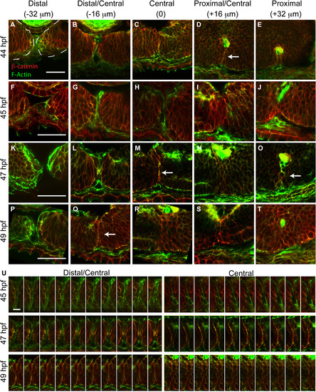
Temporal and spatial dynamics of tissue fusion during choroid fissure closure in zebrafish. Single micron optical sections from sagittal cryosections stained with phalloidin (green) and anti-β-catenin (red) at distinct proximal-distal regions of the CF over time. As in Fig. 2, the vitreous cavity was defined as central, and sections were taken at 16 µm intervals proximally and distally from this point. (A-E) At 44 hpf, the CF is fused in central/proximal sections (white arrow) based on co-localization between F-actin and β-catenin in a fusion ‘seam’. (F-J) At 45 hpf, the two sides of the CF are tightly apposed but no fusion outside of the central-proximal region is detected. (K-O) At 47 hpf, a fusion seam is present within central and proximal sections, and there are punctate regions of co-localization in distal/central sections. (P-T) At 49 hpf, the fusion seam has disappeared in the central and proximal CF regions while it is appearing in the distal/central region. (U) Single 1 µm optical sections from one section plane aligned distal (left) to proximal (right) demonstrate a progressive co-localization of F-actin and β-catenin in the CF and formation of the fusion seam. Scale bar=50 µm (A-T) and 5 µm (U). |
Reprinted from Developmental Biology, 419(2), James, A., Lee, C., Williams, A.M., Angileri, K., Lathrop, K.L., Gross, J.M., The Hyaloid Vasculature Facilitates Basement Membrane Breakdown During Choroid Fissure Closure in the Zebrafish Eye, 262-272, Copyright (2016) with permission from Elsevier. Full text @ Dev. Biol.