
Fig. 2
- ID
- ZDB-FIG-160906-3
- Publication
- Pipalia et al., 2016 - Cellular dynamics of regeneration reveals role of two distinct Pax7 stem cell populations in larval zebrafish muscle repair
- Other Figures
- All Figure Page
- Back to All Figure Page
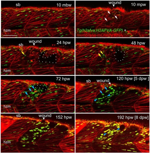
Muscle fibre regeneration in confocal time-lapse microscopy. Larvae from the Tg(h2afva:H2AFVA-GFP)kca66 line injected with membrane-mCherry RNA were wounded in epaxial somite 17 at 3.5dpf and imaged by 3D confocal time-lapse microscopy for 200hpw at 22°C. Parasagittal views are single optical slices at indicated time points from the full time series (see Movie 1). Disruption of fibres is evident immediately after wounding (white arrows). Scan shadow cast by a melanophore migrating near the wound is outlined (white dots). After loss of elongated fibre nuclei, cells with small round nuclei accumulate in wound (yellow arrows). Photobleaching resulting from scanning is evident at later times, but abundant large nuclei are located in wounds after 48hpw (blue arrows). By 5dpw, numerous rows of bright aligned nuclei are apparent (blue arrowheads). mbw, mpw, hpw and dpw: minutes before, or minutes, hours or days post-wounding; hzm, horizontal myoseptum; sb, somite border. Scale bar: 50µm. |