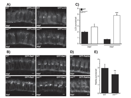
Abnormalities in nrca14 cones are specific to late endosomes and autophagosomes and appear early in development. Images of fixed wild type (WT) and nrca14 retinas from A: Tg(TαCP : GFP-map1lc3b) and B: Tg(TαCP : GFP-rab7) larvae at 3 and 4 days post-fertilization (dpf). C: nrca14 cones contain more LC3 positive puncta than WT cones by 3 dpf (compare left panels in A). B: Abnormally enlarged Rab7 structures are present in nrca14 cones by 3 dpf. D: Images of fixed WT and nrca14 retinas from 5 dpf Tg(TαCP : GFP-Rab5a) larvae. There is no difference in the appearance or number of Rab5a positive early endosomes between WT and nrca14. Scale bar = 2 µm in all images. Graph C shows average LC3 puncta per cell at 3 or 4 dpf, error bars are standard error of mean (SEM). n = 8 WT larvae and seven nrca14 larvae on 3 dpf and n = 8 WT larvae and nine nrca14 larvae on 4 dpf. (*p-value < 0.05, ***p-value < 0.001 as assessed by Mann-Whitney test). Graph E shows average Rab5a puncta per cell at 5 dpf, error bars are SEM. n = 4 WT larvae and four nrca14 larvae. (ns = p-value > 0.05 as assessed by Mann–Whitney test).
|
