
Fig. 2
- ID
- ZDB-FIG-160726-37
- Publication
- Barber et al., 2016 - Similarly Lethal Strains of Extraintestinal Pathogenic Escherichia coli Trigger Markedly Diverse Host Responses in a Zebrafish Model of Sepsis
- Other Figures
- All Figure Page
- Back to All Figure Page
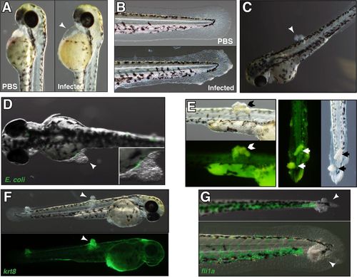
Distinct pathologies associated with different but equally lethal ExPEC isolates. (A) An F11-infected embryo displaying characteristic pericardial edema (arrowhead) at 12 hpi. This is not seen following the injection of controls with PBS (left). (B) A CFT073-infected embryo at 12 hpi showing fin erosion and ulceration commonly seen during infection with either CFT073 or F11. (C) F11-infected embryos, but not those infected with CFT073, often develop protrusions (arrowhead) in the trunk or tail region by 12 hpi. (D) Merged bright-field and fluorescent images of an F11/pGEN-GFP(LVA)-infected embryo at 12 hpi showing bacteria at the base of a protrusion (inset) but not within the main structure. (E) Bright-field and matched fluorescent images of F11-infected embryos stained with acridine orange (green) at 12 hpi. The dye accumulates within protrusions on the trunk (left, Chevron) and tail (right, arrows). (F) Bright-field (top) or fluorescent (bottom) images of Tg(krt8:GFP) embryos at 12 hpi with F11. Epithelial cells within this transgenic line express GFP. Each arrowhead marks an F11-induced protrusion. (G) Merged bright-field and fluorescent images of Tg(fli1a:GFP) embryos at 12 hpi with F11. Endothelial cells and leukocytes in this transgenic line express GFP. Protrusions are indicated by arrowheads. |
| Gene: | |
|---|---|
| Fish: | |
| Condition: | |
| Anatomical Terms: | |
| Stage: | Pec-fin |
| Fish: | |
|---|---|
| Condition: | |
| Observed In: | |
| Stage: | Pec-fin |