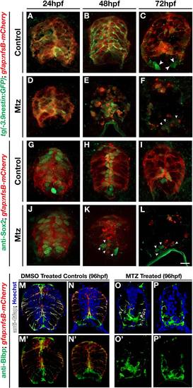
Fig. 6
- ID
- ZDB-FIG-160606-15
- Publication
- Johnson et al., 2016 - Gfap-positive radial glial cells are an essential progenitor population for later-born neurons and glia in the zebrafish spinal cord
- Other Figures
- All Figure Page
- Back to All Figure Page
|
Radial glial ablation reduces the neural progenitor population. (A-F) Cross sections of 24, 48, and 72 hpf double transgenic gfap:nfsb-mCherrysc059;-3.9nestin:GFP embryos treated with (A-C) vehicle control or (D-F) Mtz. All sections were immunolabeled with anti-GFP to enhance the signal of the 3.9nestin:gfp transgene. (G-L) Cross section views of 24, 48, and 72 hpf gfap:nfsb-mCherrysc059 embryos immunolabeled with anti-Sox2 (green) to visualize neural stem cell populations following treatment with (G-I) vehicle control or (J-L) Mtz. Ventral populations not lost following ablation are denoted (arrowheads). (M-P) Two examples of transverse sections of the spinal cord of 96 hpf gfap:nfsB-mCherry control (M, N) and Mtz-treated (O, P) embryos labeled for Blbp (green), Gfap (white), and nuclei (Hoechst). (M′-P′) mCherry and Blbp channels for the corresponding sections seen in M-P. A single Blbp+ midline cells can be seen with minute mCherry expression in a small lateral membrane process (M′ arrowhead); however, more laterally positioned Blbp cells were consistently negative for mCherry (M′, N′) as was the vast majority of Blbp labeling following gfap-mediated ablation (O′,P′). Scale bar = 10µm. |

