
Fig. S21
- ID
- ZDB-FIG-160303-35
- Publication
- Chen et al., 2016 - Efficient extravasation of tumor-repopulating cells depends on cell deformability
- Other Figures
- All Figure Page
- Back to All Figure Page
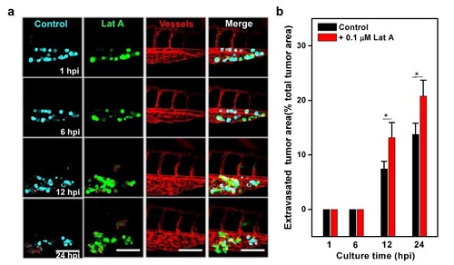
Softening shSox2 TRCs via actin depolymerization increases tumor extravasation in fish. TRCs were transfected with GFP-Sox2 shRNA (green) on the top of soft fibrin gels for 12 hrs, then treated with 0.1 µM Latrunculin A (Lat A) for 1 hr and 0.004% DMSO was used as a control. For DMSO-treated group, pECFP-N1 plasmids were co-transfected into GFP-shSox2-TRCs; for Lat A-treated group, non-fluorescent vector plasmids were co-transfected into GFP-shSox2-TRCs. Two types of tumor cells were mixed and co-injected at 1:1 ratio into the cavity of pericardium of 48 hpf embryos. (a) Representative images show tumor cells coextravasation at 1, 6, 12, and 24 hpi. Dashed red lines mark tumor extravasation areas from vessels to surrounding tissues. DMSO-treating cells (control) are cyan, Lat Atreating cells (Lat A) are green, and vessels are red. Scale bars, 100 µm. (b) Quantification of extravasated tumor areas at 1, 6, 12, and 24 hpi. Note that 0.1 µM Lat A significantly increased the extravasation of shSox2-TRCs. Mean±s.e.m.; n=18 larvae; *p < 0.05. |