
Fig. 2
- ID
- ZDB-FIG-160208-17
- Publication
- Sisson et al., 2015 - A role of glypican4 and wnt5b in chondrocyte stacking underlying craniofacial cartilage morphogenesis
- Other Figures
- All Figure Page
- Back to All Figure Page
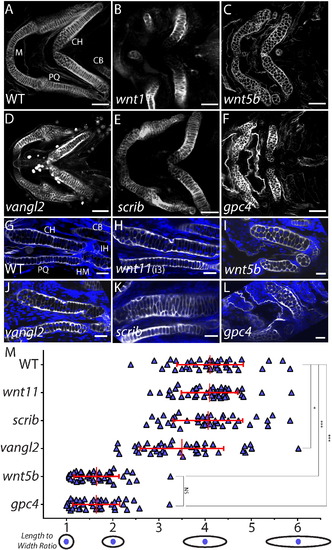
A subset of Wnt/PCP molecules influence craniofacial cartilage development. (A–F) Not all identified core Wnt/PCP zebrafish mutants have a craniofacial chondrocyte stacking defect. Confocal images of whole-mount Wheat Germ Agglutanin (WGA) staining (gray) of Wnt/PCP mutant embryo heads at 4 dpf. Scale bar = 50 µm. (G–L) Confocal images of a single focal plan of WGA staining (gray) of Wnt/PCP mutant craniofacial cartilage elements at 4 dpf. Nuclei were labeled with DAPI (blue). Scale bar = 20 µm. M. Graph of length to width ratio. The y-axis is all 40 chondrocyte measurements from 4 different ceratohyals for wild type and mutant genotypes. The x-axis is the length to width ratio with a graphical representation of select ratios. The mean LWR and their respective standard deviation is marked by red cross-hairs and stated in Supplemental Table 1. To determine if data could be compared pairwise the D′Agostino and Pearson omnibus normality test was used. Mann–Whitney U was determined and the statistical significances between the means is marked on graph and stated in Supplemental Table 1. Abbreviations: M = Meckel′s cartilage, CH = ceratohyal, CB = ceratobranchial, HM = hyomandibular, IH = interhyal joint, and PQ = palatoquadrate. (For interpretation of the references to color in this figure legend, the reader is referred to the web version of this article.) |
| Fish: | |
|---|---|
| Observed In: | |
| Stage: | Day 4 |
Reprinted from Mechanisms of Development, 138 Pt 3, Sisson, B.E., Dale, R.M., Mui, S.R., Topczewska, J.M., Topczewski, J., A role of glypican4 and wnt5b in chondrocyte stacking underlying craniofacial cartilage morphogenesis, 279-90, Copyright (2015) with permission from Elsevier. Full text @ Mech. Dev.