
Fig. 6
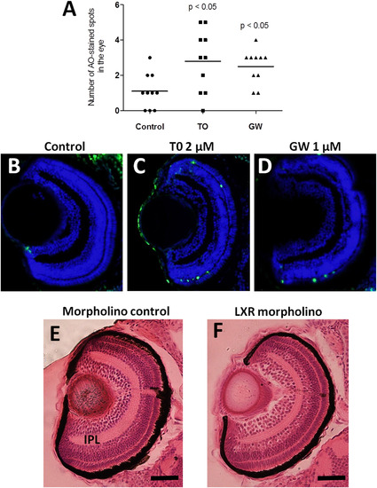
Apoptosis and morphological alterations in the eyes of Lxr ligand-treated or Lxr morpholino injected zebrafish larvae. Number of acridine orange stained spots in eyes of 4 dpf zebrafish larvae treated with T0 and GW (A). TUNEL staining of eye sections of zebrafish larvae treated vehicle only (B), T0 (C) or GW (D), Hematoxylin and eosin staining of eye sections of control morpholino (E) and Lxr morpholino (F) zebrafish larvae at 4 dpf. Representative images are shown. IPL; inner plexiform layer (Scale bars 50 µm). (For interpretation of the references to colour in this figure legend, the reader is referred to the web version of this article.) |
| Fish: | |
|---|---|
| Condition: | |
| Observed In: | |
| Stage: | Day 4 |
Reprinted from Molecular and Cellular Endocrinology, 419, Pinto, C.L., Kalasekar, S.M., McCollum, C.W., Riu, A., Jonsson, P., Lopez, J., Swindell, E., Bouhlatouf, A., Balaguer, P., Bondesson, M., Gustafsson, J.Å., Lxr regulates lipid metabolic and visual perception pathways during zebrafish development, 29-43, Copyright (2016) with permission from Elsevier. Full text @ Mol. Cell. Endocrinol.