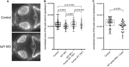
Fig. 4
- ID
- ZDB-FIG-160205-31
- Publication
- O'Rawe et al., 2015 - TAF1 Variants Are Associated with Dysmorphic Features, Intellectual Disability, and Neurological Manifestations
- Other Figures
- All Figure Page
- Back to All Figure Page
|
Suppression or Genetic Mutation of Endogenous taf1 Induces Decreased Size of the Optic Tectum In Vivo (A) Dorsal view of a control embryo (top) and an embryo injected with a morpholino (MO) targeting the donor site of exon 9 of D. rerio taf1 3 days after fertilization. An antibody against α-acetylated tubulin was used for visualizing the axon tracts in the brain of evaluated embryos. The assay consisted of measuring the area of the optic tectum (highlighted with the dashed ellipse), a neuroanatomical structure that occupies the majority of the space within the midbrain. (B) A boxplot shows quantitative differences in the size of the optic tectum for each condition tested across three biological replicates. Suppression of taf1 consistently induced a decrease of ~10% in the relative area of the optic tectum (p < 0.0001). The MO phenotype could be restored by co-injection of MO and wild-type (WT) human TAF1 mRNA (p < 0.0001), denoting the specificity of the phenotype due to taf1 suppression. Overexpression of WT human TAF1 mRNA alone did not induce a phenotype that was significantly different from that of controls (p = 0.79). The numbers of embryos evaluated per condition were as follows: control, 134; taf1 MO, 133; taf1 MO + WT TAF1 RNA, 109; and WT TAF1 RNA, 78. (C) A boxplot shows quantitative differences in the size of the optic tectum between uninjected controls and F0 embryos with CRISPR-disrupted taf1. The phenotype observed for both MO-injected embryos and embryos with CRISPR-disrupted taf1 was concordant and reproducible across different experiments and across the two different methodologies. The p values were calculated with a Student’s t test. |
| Fish: | |
|---|---|
| Knockdown Reagents: | |
| Observed In: | |
| Stage: | Protruding-mouth |

