
Fig. 6
- ID
- ZDB-FIG-151113-16
- Publication
- Kimura et al., 2015 - Integration of vascular Systems between the brain and spinal cord in zebrafish
- Other Figures
- (all 7)
- All Figure Page
- Back to All Figure Page
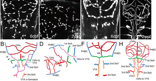
Remodeling process for the temporal integration of cerebral and spinal vascular systems. Multi-photon microscopic image of Tg(fli1a:EGFP)y7 embryos at 6 (A and E) and 7 dpf (C); microangiography with highlighter ink of casper larvae at 2.5 wpf (G); and their schematic diagrams (B, D, F and H). Dorsal (A, E, and G) and dorso-lateral (C) views. Two pathways, connecting vascular systems, are indicated by colored lines (B, D and H pink lines: supplemental route of CtAs to VTAs; B, F and H brown lines: conventional route of BA-1st SeAs to VTAs). Arrows in A and B indicate the disconnection of the PHBC and DLAV. |
Reprinted from Developmental Biology, 406(1), Kimura, E., Isogai, S., Hitomi, J., Integration of vascular Systems between the brain and spinal cord in zebrafish, 40-51, Copyright (2015) with permission from Elsevier. Full text @ Dev. Biol.