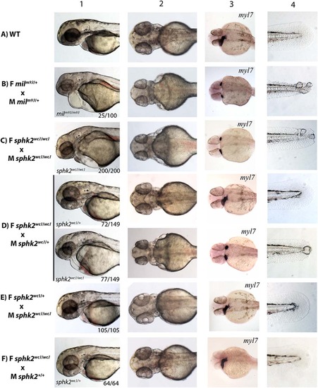
The sphk2MZ mutant phenocopies the mil zygotic mutant. Shown are representative embryos at 32 hr post fertilization imaged in brightfield (column 1: lateral view, anterior to the left), (column 2: ventral view, anterior to the left), or analyzed by in situ hybridization for expression of the myl7 gene (column 3, ventral view, anterior to the left). Column 4 shows representative images of tails at this stage. Embryos were derived from crosses as indicated on the left: (A) Wildtype (WT), (B) heterozygous mil adults, (C) female and male sphk2wc1/wc1 mutants, (D) female sphk2 homozygous mutant and male sphk2 heterozygous mutant and (E) female sphk2 heterozygous mutant and male sphk2 homozygous mutant. (F) Female sphk2 homozygous mutant crossed to a wildtype male. Note that only the homozygous mil mutant embryos (25% of the embryos from the heterozygote parental cross) display pericardial edema (columns 1, 2), cardia bifida (column 3), and tail blisters (column 4), as do 100% (200/200) of the sphk2MZ mutant embryos. These phenotypes are rescued in embryos that have a single wildtype allele (72/149, 48% of the embryos in D), if embryos inherit maternal message from a wildtype maternal allele (50% of the embryos in E, which are zygotically null but phenotypically normal), or if embryos inherit a single wildtype allele from the male parent (64/64, 100% of the embryos in F).
|
