
Fig. 3
- ID
- ZDB-FIG-151002-41
- Publication
- Riemer et al., 2015 - A functional Bucky ball-GFP transgene visualizes germ plasm in living zebrafish
- Other Figures
- All Figure Page
- Back to All Figure Page
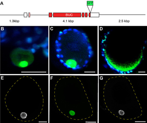
Buc-GFP transgenic zebrafish. (A) Scheme showing the genomic Buc locus used to generate the Buc-GFP line. Thin lines represent non-coding regions. Boxes represent exons (white: untranslated RNA; red: coding RNA). The green box marks the insertion point of the GFP-protein in front of the STOP-codon. (B–D) Live images of Buc-GFP (green) in oocytes from transgenic mothers at early stage IB (B), late IB (C), and II (D). The nuclei of follicle cells were counterstained with the live-cell fluorescent dye Hoechst 33342 (blue). (E–G) Confocal images comparing endogenous Buc localization with Buc-GFP. (E) Wild-type stage IB oocyte labeled with Buc antibody (white) compared to transgenic Buc-GFP oocyte labeled with GFP antibody (F; green) and Buc (G; white). Dashed yellow line indicates oocyte outline (E–G). Lateral view, animal to the top. Scale bar: 10 µm (E–G) 25 µm (B–D). |
| Gene: | |
|---|---|
| Fish: | |
| Anatomical Terms: | |
| Stage: | Adult |
Reprinted from Gene expression patterns : GEP, 18(1-2), Riemer, S., Bontems, F., Krishnakumar, P., Gömann, J., Dosch, R., A functional Bucky ball-GFP transgene visualizes germ plasm in living zebrafish, 44-52, Copyright (2015) with permission from Elsevier. Full text @ Gene Expr. Patterns