Fig. S4
- ID
- ZDB-FIG-150921-35
- Publication
- Anderson et al., 2015 - In vivo Modeling Implicates APOL1 in Nephropathy: Evidence for Dominant Negative Effects and Epistasis under Anemic Stress
- Other Figures
- All Figure Page
- Back to All Figure Page
|
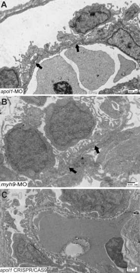
Further characterization of apol1 and myh9 morphant glomerular ultrastructure. Transmission electron microscopy of zebrafish larval glomeruli injected with either (A) apol1-MO or (B) myh9-MO were imaged at 5 dpf using a low magnification (direct mag = 4400X) to characterize long stretches of the glomerular basement membrane (GBM). Comparatively, apol1 and myh9 morphants display similar abnormalities, including podocyte disorganization and effacement, as well as the presence of microvillus protrusions. However, myh9 morphants display a thickened GBM that is not apparent in apol1-MO injected larvae, while apol1 morphants appear to have a higher degree of podocyte effacement compared to myh9 morphants. (C) Zebrafish larvae injected with apol1 CRISPR/CAS9 display a similar glomerular ultrastructure compared to apol1 morphants at 5 dpf. Filled arrowheads, glomerular basement membrane. Scale bar = 500nm. |
| Fish: | |
|---|---|
| Knockdown Reagents: | |
| Observed In: | |
| Stage: | Day 5 |

