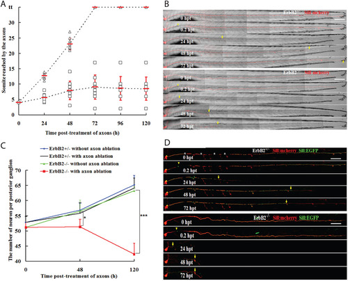
Schwann cells facilitate but are dispensable for axonal regeneration. (A) The axons were severed at somite 3 in 5-dpf larvae. The somite reached by the regenerating nerve was examined at 24, 48, 72, 96 and 120hpt. In all cases (n=10), the axons had reached the tip of the body and re-innervated the terminal neuromasts at 72hpt in Tg[gSAGFF202A+/SILL:mCherry] (triangles). All cases (n=10) of Tg[gSAGFF202A/SILL:mCherry] fish revealed an abnormal and lower regeneration rate (squares). (B) Examples of axon regeneration after injury in heterozygous and homozygous Tg[gSAGFF202A;SILL:mCherry]. (C) Sum of neuron numbers in posterior ganglion at 5dpf, 48hpt and 120hpt in Tg[gSAGFF202A+/SILL:mCherry] and Tg[gSAGFF202A/SILL:mCherry] larvae upon laser-mediated axon severing or in the untreated group. n=6 fish per group (*P<0.05, ***P<0.0001). (D) Examples of an identified axon’s regeneration after damage in heterozygous and homozygous Tg[gSAGFF202A;SILL:mCherry] fish injected with SILL:EGFP. Yellow arrowheads and arrows point to cutting sites and to the tip of regenerate axons, respectively. White asterisks mark pigment cells. Error bars are+or±s.d. Scale bars: 150µm (B,D).
|
