
Fig. 1
- ID
- ZDB-FIG-150421-4
- Publication
- Navis et al., 2015 - Loss of cftr function leads to pancreatic destruction in larval zebrafish
- Other Figures
- All Figure Page
- Back to All Figure Page
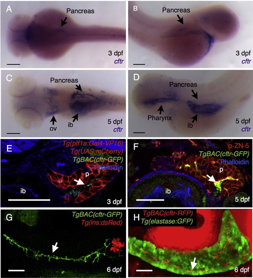
cftr is expressed in the pancreatic duct. (A–D) Wholemount in situ hybridization for cftr. (A) Dorsal and (B) lateral view of cftr expression at 3 dpf. (C) Dorsal and (D) lateral view of cftr expression at 5 dpf. (E and F) Transverse section of TgBAC(cftr-GFP) expressed in the pancreatic duct at (E) 3 dpf in conjunction with ptf1a expression in the acinar cells of the pancreas and (F) at 5 dpf in conjunction with ZN-5, an antibody that marks the pancreas. (G) Live, confocal image of TgBAC(cftr-GFP) expression along the length of the pancreatic duct in conjunction with Tg(ins:dsRed) to mark the β cells of the principal islet at 5 dpf. (H) Live confocal image of ductal TgBAC(cftr-RFP) expression in conjunction with ela:GFP to indicate the acinar tissue. White arrows indicate cftr expression in the pancreatic duct. ib: intestinal bulb, ov: otic vesicle, p: pancreas. (A–D) Scale bars=100 µm, (E–H) Scale bars=50 µm. |
| Genes: | |
|---|---|
| Antibody: | |
| Fish: | |
| Anatomical Terms: | |
| Stage Range: | Protruding-mouth to Day 6 |
Reprinted from Developmental Biology, 399(2), Navis, A., Bagnat, M., Loss of cftr function leads to pancreatic destruction in larval zebrafish, 237-48, Copyright (2015) with permission from Elsevier. Full text @ Dev. Biol.