|
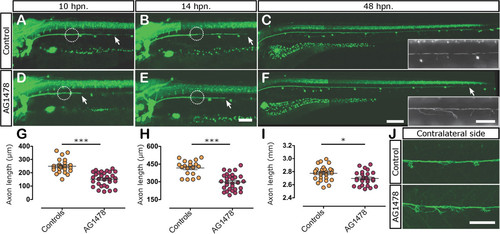
Role of ErbB signaling in axonal regeneration. Three days post fertilization larvae were neurectomized (circles indicate position of neurectomy) after incubation with 0.05% of DMSO (controls) or 5 µM of AG1478 from 60 hpf to 5 dpf (48 hpn); this treatment does not affect Schwann cell survival but blocks ErbB signaling during the regeneration phase. Nerve regeneration in control (A-C) and drug treated larvae (D-F) was evaluated after 10 hpn, 14 hpn, and 48 hpn. The insets in C and F show the defasciculation of the regenerated nerve in treated larvae in comparison to controls. (G-I) Quantitative analysis of axonal extension after 10 hpn (G), 14 hpn (H), and 48 hpn (I) in control and AG1478 treated larvae. *P <0.05; ***P <0.001. (J) In 30% of ErbB inhibitor treated larvae, the non-neurectomized contralateral nerve appears defasciculated in comparison to the control nerve. Scale bar: C, F: 200 µm; A, B, D, E, J; insets in C and F: 100 µm.
|

